Template:Infobox drug mechanism/testcases
| This is the template test cases page for the sandbox of Template:Infobox drug mechanism. to update the examples. If there are many examples of a complicated template, later ones may break due to limits in MediaWiki; see the HTML comment "NewPP limit report" in the rendered page. You can also use Special:ExpandTemplates to examine the results of template uses. You can test how this page looks in the different skins and parsers with these links: |
| {{Infobox drug mechanism}} | {{Infobox drug mechanism/sandbox}} | ||||||||||||||||||||||||||||||||||||||||
|---|---|---|---|---|---|---|---|---|---|---|---|---|---|---|---|---|---|---|---|---|---|---|---|---|---|---|---|---|---|---|---|---|---|---|---|---|---|---|---|---|---|
|
| ||||||||||||||||||||||||||||||||||||||||
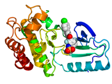
- ^ PDB: 3OG7; Bollag G; Hirth P; et al. (September 2010). "Clinical efficacy of a RAF inhibitor needs broad target blockade in BRAF-mutant melanoma". Nature. 467 (7315): 596–599. doi:10.1038/nature09454. PMC 2948082. PMID 20823850.
{{cite journal}}: Unknown parameter|author-separator=ignored (help)

